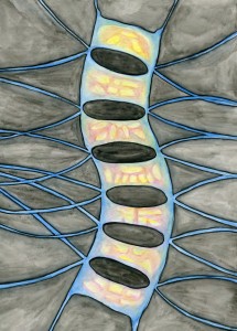
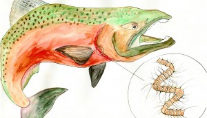
When viewing a chaetoceros with a compound microscope under 100x, it appears as a series of rectangular cells, connected by interweaving spines. If viewed with an electron microscope, they are elliptical in appearance when observed in valve view. They have two spines arising from each valve. Adjacent cells are connected by crossing of the spines without direct contact of the cell body. (Cupp 1943). The species with larger, thicker spines can cause physical damage to organisms’ gills when present in large numbers (Kraberg et al. 2010). Chaetoceros are found in the neritic zone, making them a potential threat to hatchery fish that live along coastlines. (Cupp 1943) These diatoms are harmless to humans but have the potential to wreak havoc on overcrowded hatchery fish. The increased amount of nutrients that are being provided for the fish, as well as from their excrement can cause harmful algal blooms. This process is called eutrophication and chaetoceros with large spines are a particular threat to fish, as they get caught in their gills.
Kingdom: Chromista
Phylum: Bacillariophyta
Class: Mediophyceae
Subclass: Chaetocerotophycidae
Order: Chaetocerotales
Family: Chaetocerotaceae


